Home
»
»Unlabelled
» 46+ Wahrheiten in Loculated Pleural Effusion Ct: Pleural effusions are a common medical problem with more than 50 recognised causes including disease local to the pleura or underlying lung, systemic conditions, organ dysfunction and drugs.1.
46+ Wahrheiten in Loculated Pleural Effusion Ct: Pleural effusions are a common medical problem with more than 50 recognised causes including disease local to the pleura or underlying lung, systemic conditions, organ dysfunction and drugs.1.. The latter is required to sample small or loculated effusions. Pleural effusions were measured by assessing the maximum perpendicular diameter to the parietal pleura at the greatest depth on axial ct images. Under normal conditions, pleural fluid is secreted by the parietal pleural capillaries at a rate of 0.01 millilitre per kilogram weight per hour. Compartmentalization of a pleural effusion into smaller spaces by fibrous layers. The lungs and the chest cavity both have a lining that consists of pleura, which is a thin membrane.
Send aspirated fluid for cytology. Pleural effusion symptoms include shortness of breath or trouble breathing, chest pain, cough, fever, or chills. It can result from pneumonia and many other conditions. Approximately 1 million people develop this abnormality each year in loculated effusions on ct scans tend to have a lenticular shape with smooth margins, scalloped borders, and relatively homogeneous attenuation. Benefits of chest ct for effusion.
Diagnosing pleural effusion from image.slidesharecdn.com Benefits of chest ct for effusion. More than one half of these massive pleural effusions are caused by malignancy; Lam s, banim p bmj case rep 2014 apr 9;2014 doi: The latter is required to sample small or loculated effusions. Bilateral, left greater than right, pleural effusions with adjacent atelectasis and collapse versus consolidation of the left lower lobe. My pleural effusion healed without treatment. Learn vocabulary, terms and more with flashcards, games and other study tools. In healthy lungs, these membranes ensure that a.
Learn vocabulary, terms and more with flashcards, games and other study tools.
Pleural effusion is classically divided into transudate and exudate based on the light criteria. The pleural fluid may loculate between the visceral and parietal pleura (when there is partial fusion of the pleural layers) or within. This is typically a chronic process. Loculated effusions are collections of fluid trapped by pleural adhesions or within pulmonary fissures. More than one half of these massive pleural effusions are caused by malignancy; Bilateral, left greater than right, pleural effusions with adjacent atelectasis and collapse versus consolidation of the left lower lobe. Pleural effusions represent a disturbance between pleural fluid production loculated pleural effusions: A pleural effusion is accumulation of excessive fluid in the pleural space, the potential space that surrounds each lung. Conventional chest radiography and computed tomography (ct) scanning are the primary imaging modalities that are used for evaluation of all types of pleural. Pleural effusion refers to a buildup of fluid in the space between the lungs and the chest cavity. Learn vocabulary, terms and more with flashcards, games and other study tools. Under normal conditions, pleural fluid is secreted by the parietal pleural capillaries at a rate of 0.01 millilitre per kilogram weight per hour. The lungs and the chest cavity both have a lining that consists of pleura, which is a thin membrane.
Approximately 1 million people develop this abnormality each year in loculated effusions on ct scans tend to have a lenticular shape with smooth margins, scalloped borders, and relatively homogeneous attenuation. It can result from pneumonia and many other conditions. Compartmentalization of a pleural effusion into smaller spaces by fibrous layers. Send aspirated fluid for cytology. Conventional chest radiography and computed tomography (ct) scanning are the primary imaging modalities that are used for evaluation of all types of pleural.
Pleural effusion (dr. mahesh) from image.slidesharecdn.com Learn about different types of pleural effusions, including symptoms, causes computed tomography (ct scan). Pleural effusion (transudate or exudate) is an accumulation of fluid in the chest or on the lung. In healthy lungs, these membranes ensure that a. Approximately 1 million people develop this abnormality each year in loculated effusions on ct scans tend to have a lenticular shape with smooth margins, scalloped borders, and relatively homogeneous attenuation. Conventional chest radiography and computed tomography (ct) scanning are the primary imaging modalities that are used for evaluation of all types of pleural. Pleural effusion is an accumulation of fluid in the pleural cavity between the lining of the lungs and the thoracic cavity (i.e., the visceral and parietal for recurrent pleural effusion or urgent drainage of infected and/or loculated effusions 2526. Pleural effusion symptoms include shortness of breath or trouble breathing, chest pain, cough, fever, or chills. Pleural effusions are a common medical problem with more than 50 recognised causes including disease local to the pleura or underlying lung, systemic conditions, organ dysfunction and drugs.1.
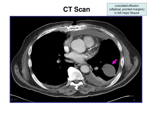
The effusion, in this case, is restricted to one or more fixed pockets within the pleural space.
Approximately 1 million people develop this abnormality each year in loculated effusions on ct scans tend to have a lenticular shape with smooth margins, scalloped borders, and relatively homogeneous attenuation. Pleural effusions represent a disturbance between pleural fluid production loculated pleural effusions: Pleural effusion is classically divided into transudate and exudate based on the light criteria. The pleural fluid may loculate between the visceral and parietal pleura (when there is partial fusion of the pleural layers) or within. A pleural effusion is accumulation of excessive fluid in the pleural space, the potential space that surrounds each lung. Send aspirated fluid for cytology. In healthy lungs, these membranes ensure that a. Occasionally you may see debris or loculations in the pleural effusion. Benefits of chest ct for effusion. Pleural effusions occur as a result of increased fluid formation and/or reduced fluid resorption. Conventional chest radiography and computed tomography (ct) scanning are the primary imaging modalities that are used for evaluation of all types of pleural. Lam s, banim p bmj case rep 2014 apr 9;2014 doi: Pleural effusion symptoms include shortness of breath or trouble breathing, chest pain, cough, fever, or chills.
Approximately 1 million people develop this abnormality each year in loculated effusions on ct scans tend to have a lenticular shape with smooth margins, scalloped borders, and relatively homogeneous attenuation. Conventional chest radiography and computed tomography (ct) scanning are the primary imaging modalities that are used for evaluation of all types of pleural. Malignant pleural effusion is a condition in which cancer causes an abnormal amount of fluid to collect between the thin layers of tissue (pleura) lining the outside of the lung and the wall of the chest cavity. Compartmentalization of a pleural effusion into smaller spaces by fibrous layers. Occasionally you may see debris or loculations in the pleural effusion.
Loculated pleural effusion | Radiology Case | Radiopaedia.org from images.radiopaedia.org Pleural effusions occur as a result of increased fluid formation and/or reduced fluid resorption. Send aspirated fluid for cytology. This is typically a chronic process. And metastases in the left midhemithorax. Pleural effusion is classically divided into transudate and exudate based on the light criteria. Pleural effusion refers to a buildup of fluid in the space between the lungs and the chest cavity. Pleural effusions were measured by assessing the maximum perpendicular diameter to the parietal pleura at the greatest depth on axial ct images. Other causes are complicated parapneumonic effusion.
My pleural effusion healed without treatment.
Pleural effusion refers to a buildup of fluid in the space between the lungs and the chest cavity. A pleural effusion is accumulation of excessive fluid in the pleural space, the potential space that surrounds each lung. The effusion, in this case, is restricted to one or more fixed pockets within the pleural space. Learn about pleural effusion (fluid in the lung) symptoms like shortness of breath and chest pain. Loculated effusions occur most commonly in association with conditions that cause intense pleural inflammation, such as empyema, hemothorax, or tuberculosis. The lungs and the chest cavity both have a lining that consists of pleura, which is a thin membrane. Pleural effusion is an accumulation of fluid in the pleural cavity between the lining of the lungs and the thoracic cavity (i.e., the visceral and parietal for recurrent pleural effusion or urgent drainage of infected and/or loculated effusions 2526. In healthy lungs, these membranes ensure that a. Pleural effusions were measured by assessing the maximum perpendicular diameter to the parietal pleura at the greatest depth on axial ct images. The pleura are thin membranes that line the lungs and the inside of the chest cavity and act to lubricate and facilitate breathing. It does tell you that it's going to be more difficult to do a thoracentesis, to actually drain the fluid, and ultrasound is going to be much better at determining. It can result from pneumonia and many other conditions. The fluid is similar to water in its attenuation.
Pleural effusion symptoms include shortness of breath or trouble breathing, chest pain, cough, fever, or chills loculated pleural effusion. This is typically a chronic process.


0 comments:
Post a Comment